Pelvic Anatomy Female Ligaments / Female Pelvis with Ligaments, midsagitally sectioned ... : Functional anatomy of the male pelvic floor online course:
byAdmin•
0
Pelvic Anatomy Female Ligaments / Female Pelvis with Ligaments, midsagitally sectioned ... : Functional anatomy of the male pelvic floor online course:. Of female pelvic organ sacrospinous ligament just medial to the ischial spine, exiting the pelvis through the greater sciatic foramen. These ligaments arise from the side of the cervix and the lateral fornix of the vagina. Ischial tuberosities, sacrotuberous and sacrospinous ligaments and, tip of the coccyx. Above the pelvic brim and has no obstetric importance. 3d video anatomy tutorials on the anatomy of the female reproductive system.
Learn now at kenhub their anatomy! Related online courses on physioplus. This anatomy section promotes the use of the terminologia anatomica, the international standard of anatomical nomenclature. Abdominal and pelvic anatomy encompasses the anatomy of all structures of the abdominal and pelvic cavities. They provide an extensive attachment on the lateral pelvic wall at the level of the ischial spines.
The Pelvic Girdle and Pelvis | Anatomy and Physiology I from s3-us-west-2.amazonaws.com In front it is incomplete, presenting a wide interval between the anterior borders of the ilia. Raz s, rodriguez l, editors. Learn now at kenhub their anatomy! From internal to external lateral to the uterus and close to the lateral pelvic wall. Formulary drug information for this topic. Fascia which lines the pelvic cavity, viscera and vessels condenses to form ligaments: Abdominal and pelvic anatomy encompasses the anatomy of all structures of the abdominal and pelvic cavities. The female bony pelvis is divided into:
The broad ligament is related to many structures within the female pelvis.
Raz s, rodriguez l, editors. Pelvic anatomy sacrouterine ligament cardinal ligaments pelvic fascia sacrospinous ligament urethral support bladder support rectal support. In females, the pelvis also houses the uterus, fallopian tubes, and ovaries. The female bony pelvis is divided into: Formulary drug information for this topic. Peritoneum and the broad ligament. Mr assessment of variations during the. Fascia which lines the pelvic cavity, viscera and vessels condenses to form ligaments: Ischial tuberosities, sacrotuberous and sacrospinous ligaments and, tip of the coccyx. Learn vocabulary, terms and more with flashcards, games and other study tools. • divided into the true and false pelvis by the iliopectineal continuation of the broad ligament extends across the pelvic floor attaches at the isthmus portion of the uterus firmly supports the cervix. Sagittal section female pelvis with peritoneum. • pelvis begins at the iliac crests and ends at the symphysis pubis.
ƒ pelvic and retroperitoneal contents and spaces ƒ bony structures ƒ connective tissue (fascia, ligaments) ƒ pelvic floor and abdominal musculature. Four ligaments inguinal ligament • important for repair of inguial hernia cooper's ligament • frequently used in bladder suspension. 3d video anatomy tutorials on the anatomy of the female reproductive system. Suspended in the mesovarium (attached to the posterior part of the broad ligament). Functional anatomy and pathophysiology of pelvic organ prolapse.
Anatomy Model Female Pelvis Ligaments Vessels Floor from cdn11.bigcommerce.com • pelvis begins at the iliac crests and ends at the symphysis pubis. Suspended in the mesovarium (attached to the posterior part of the broad ligament). Mons pubis is a pad of fatty tissue situated. They provide an extensive attachment on the lateral pelvic wall at the level of the ischial spines. Knowledge of anatomy unique to females is essential for all clinicians three ligaments anchor the uterus. Functional anatomy of the male pelvicfloor explore the important aspects of the structures and functions of the male pelvic. Pelvic surgery requires a comprehensive knowledge of the pelvic anatomy to safely attain access, maximize exposure, ensure hemostasis, and avoid injury to viscera, blood vessels, and nerves. Ligaments and anatomy important in pelvic.
These ligaments arise from the side of the cervix and the lateral fornix of the vagina.
Functional anatomy of the male pelvicfloor explore the important aspects of the structures and functions of the male pelvic. It then enters the ischiorectal fossa through the lesser. Ischial tuberosities, sacrotuberous and sacrospinous ligaments and, tip of the coccyx. Pelvic anatomy sacrouterine ligament cardinal ligaments pelvic fascia sacrospinous ligament urethral support bladder support rectal support. Abdominal and pelvic anatomy encompasses the anatomy of all structures of the abdominal and pelvic cavities. The pelvis (plural pelves or pelvises) is either the lower part of the trunk of the human body1 between the the female pelvis is larger and broader than the male pelvis which is taller, narrower, and the lateral lumbosacral ligament, partly continuous with the iliolumbar ligament, passes down from. Knowledge of anatomy unique to females is essential for all clinicians three ligaments anchor the uterus. The uterosacral ligament supports the uterus posteriorly, and the pubocervical ligament anchors the uterus anteriorly. Vides a discussion of the contemporary understanding. Sagittal section female pelvis with peritoneum. Four ligaments inguinal ligament • important for repair of inguial hernia cooper's ligament • frequently used in bladder suspension. Peritoneum and the broad ligament. In females, the pelvis also houses the uterus, fallopian tubes, and ovaries.
These ligaments arise from the side of the cervix and the lateral fornix of the vagina. Formulary drug information for this topic. Broad ligament round ligament mesovarium mesosalpinx cardinal. This is a patient education video on normal female pelvic anatomy.www.melakafertility.com. Suspensory ligament of left ovary;
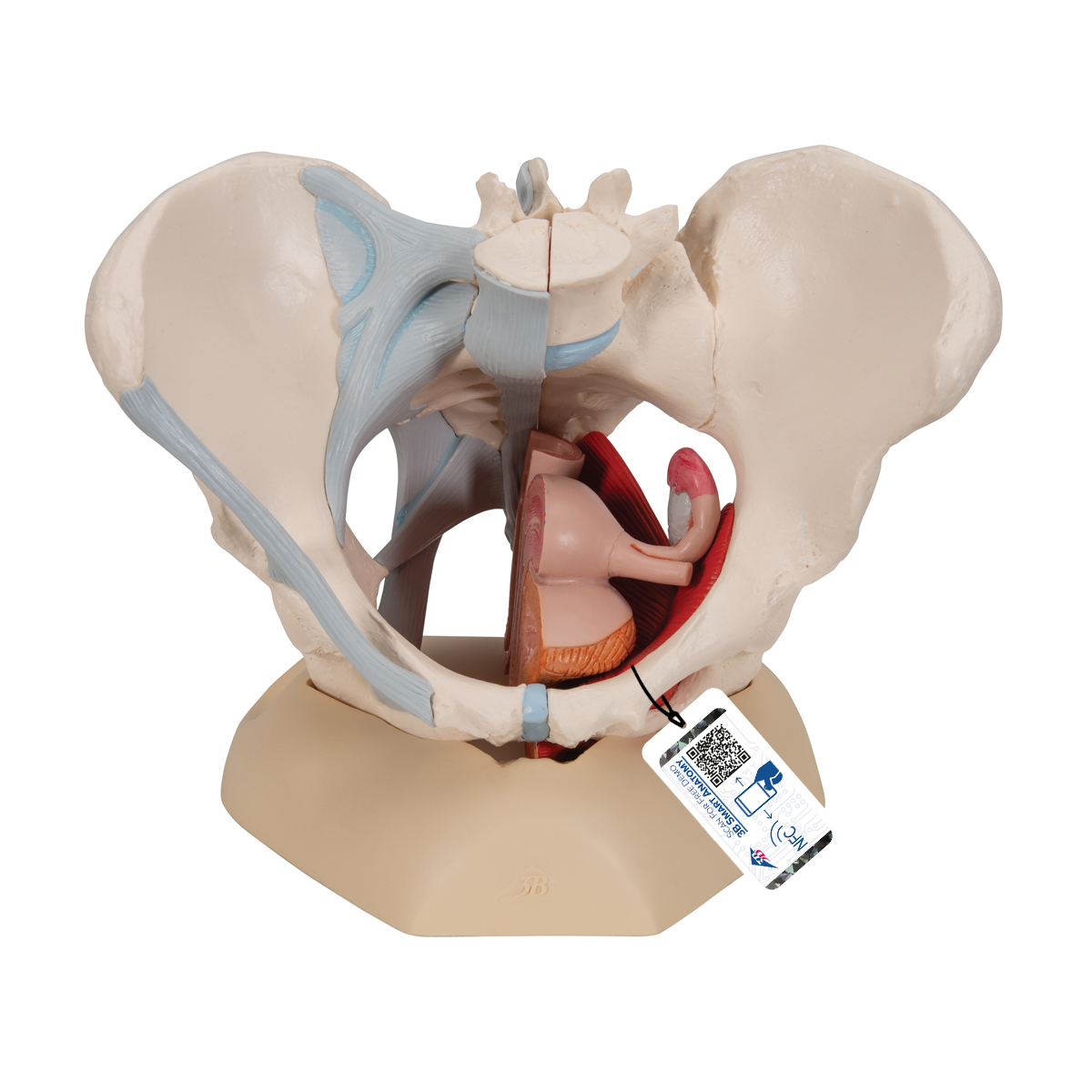
Anatomical Teaching Models - Plastic Human Pelvic Models ... from www.3bscientific.com Pelvic surgery requires a comprehensive knowledge of the pelvic anatomy to safely attain access, maximize exposure, ensure hemostasis, and avoid injury to viscera, blood vessels, and nerves. In front it is incomplete, presenting a wide interval between the anterior borders of the ilia. Four ligaments inguinal ligament • important for repair of inguial hernia cooper's ligament • frequently used in bladder suspension. The fallopian tubes are made up of three layers. Raz s, rodriguez l, editors. Mons pubis is a pad of fatty tissue situated. • pelvis begins at the iliac crests and ends at the symphysis pubis. The female bony pelvis is divided into:
• pelvis begins at the iliac crests and ends at the symphysis pubis. Formulary drug information for this topic. Peritoneum and the broad ligament. Sagittal section female pelvis with peritoneum. Vides a discussion of the contemporary understanding. Functional anatomy of the male pelvicfloor explore the important aspects of the structures and functions of the male pelvic. Suspensory ligament of left ovary; Anatomy of the pelvic floor. • divided into the true and false pelvis by the iliopectineal continuation of the broad ligament extends across the pelvic floor attaches at the isthmus portion of the uterus firmly supports the cervix. The greater or false pelvis (pelvis major).—the greater pelvis is the expanded portion of the cavity situated above and in front of the pelvic brim. Related online courses on physioplus. 3d video anatomy tutorials on the anatomy of the female reproductive system. In front it is incomplete, presenting a wide interval between the anterior borders of the ilia.
Formulary drug information for this topic pelvic anatomy. Mr assessment of variations during the.